
Part 20: Schizophrenia
Searching for a Chief Suspect

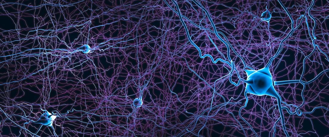
The dynamical complexity of brain circuits is daunting. Brain disorders with specific failure modes may give us insights into the normal function of specific brain circuits. For example, loss of dopamine-producing neurons in the midbrain causes Parkinson’s disease: Tremors, muscle rigidity, slow movements, and impaired balance. Thought disorders are even more enigmatic than motor disorders.
Schizophrenia is a devastating thought disorder for which we do not have effective treatments. Symptoms include: Delusions: hearing voices that do not exist, Disorganized thinking: Jumbled speech, switching topics mid-sentence, or saying things that make no sense; Flattened affect: Showing little or no emotion in the face or voice tone; Lack of drive: Trouble starting or finishing planned tasks and daily goals; Social withdrawal: Pulling away from friends, family, and group activities; Loss of pleasure: Finding no joy in life or everyday hobbies. Memory dysfunction: Difficulty using recent information or recalling details.
If we could trace the origin of these symptoms to dysfunction in a specific type of neuron or neural circuit, similar to that of dopamine neurons in Parkinson’s disease, it might be possible to design more effective treatments and unravel some of the deepest mysteries of thinking and cognitive processing. We need to find a chief suspect.
Similar symptoms appear after serial use of a party drug, Special K. Special K is ketamine, an anesthetic at high doses. At low doses, ketamine induces hallucinations, dissociative out-of-body experiences, and reduced gamma bursts, which are high-frequency bursts of spikes lasting less than a second. After two days of raving under the influence of Special K, ravers present at emergency rooms with symptoms indistinguishable from psychosis in schizophrenia patients, who also have reduced gamma bursts. Fortunately, the symptoms in the ravers go away a week later.
My lab at the Salk Institute worked on a mouse model for ketamine-induced psychosis (Behrens and Sejnowski, 2009). We discovered that ketamine downregulates GAD67, the enzyme that synthesizes the inhibitory neurotransmitter GABA in parvalbumin (PV) basket cells (Fig. 1), and also reduces gamma bursts in mice. PV basket cells make multiple “basket-like” synapses that cover the somas of neighboring pyramidal cells. Corroborating evidence comes from postmortem studies on the brains of schizophrenia patients, which revealed that their cortical PV basket cells also had lower levels of GAD67, the enzyme that synthesizes GABA, consistent with what we found in mice (Fig. 2) (Curley and Lewis, 2012). But in schizophrenia patients, the downregulation is permanent.
One of the consequences of reduced inhibition is that cortical circuits become hyperactive, which could explain hallucinations. Ketamine is also effective at relieving clinical depression, perhaps by boosting reduced cortical activity to normal levels.
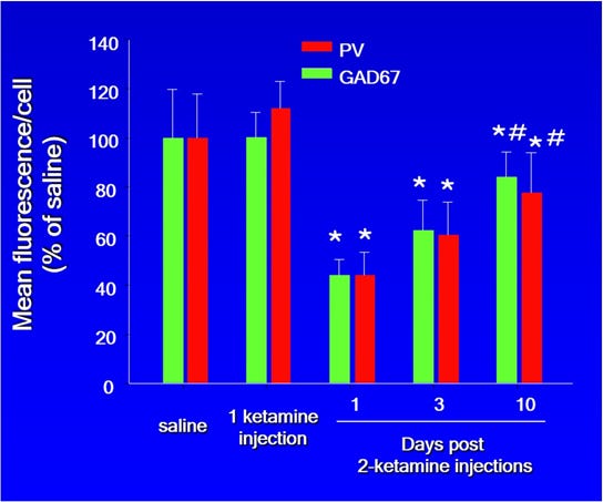
Figure 1. Mouse model of ketamine-induced psychosis. A single injection of ketamine did not change the levels of parvalbumin (PV) or GAD67 in rodent cortical PV basket cells. There was a significant decrease in both proteins following a second dose on the next day, with a gradual recovery mirroring that in humans. (Courtesy of M. Behrens)
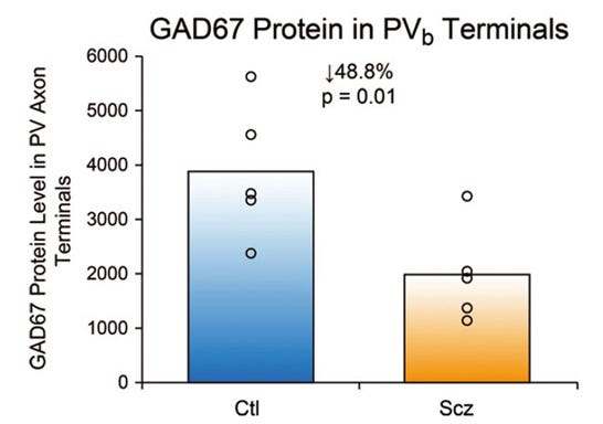
Figure 2. Postmortem schizophrenia brain (Scz) measurements of GAD67, the enzyme that synthesizes GABA, in the synaptic terminals of parvalbumin-positive basket cells (PVb). (Adapted from Curley and Lewis, 2012)
PV basket cells are the most abundant inhibitory interneurons in the neocortex. They are special because they are “fast-spiking”—they have thin spikes and can fire action potentials in bursts up to 100 Hz. Because of their bursty, high-frequency firing, basket cells are metabolically demanding and highly vulnerable to mitochondrial stress, oxidative DNA damage, and activity-induced double-strand breaks. Unlike most other inhibitory neurons, PV basket cell axons are wrapped in myelin, allowing for ultra-fast signal conduction and strict synchronization across cortical networks.
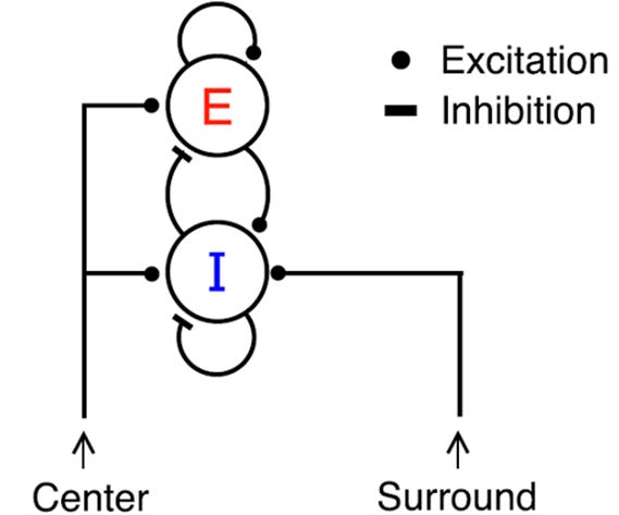
Figure 3. Schematic of the cortical circuit that is responsible for gamma bursts (Jadi and Sejnowski, 2014). The Center pathway carries direct sensory signals to excitatory pyramidal neurons (E) and inhibitory PV basket cells (I). The Surround pathway from neighboring regions of the cortex modulates the responses to sensory inputs. Each synapse can be either excitatory or inhibitory, as indicated by the shape of the contact with the postsynaptic neuron.
You are more likely to remember something the next day if you pay attention to it. Gamma bursts are generated by reciprocal connections between PV basket cells and pyramidal neurons via a ping-pong mechanism (Fig. 3) (Cardin et al., 2009; Jadi, Behrens, Sejnowski, 2016). By creating strong inhibitory inputs near the spike-initiating zone in pyramidal neurons, they exert precise, millisecond-level control over gamma bursts, which control attention and working memory. Attention changes the state of the local circuit, and gamma bursts imprint a trace of the attended stimulus into the circuit.
Schizophrenia may be a consequence of the reduced inhibition in cortical PV basket cells, just as the loss of dopamine cells causes Parkinson’s disease. Drugs to enhance dopamine activity and deep brain stimulation have ameliorated the symptoms of Parkinson’s disease. Further research led to a theory for how dopamine cells modulate motivation and broadcast reward prediction errors for reward learning (Montague, Dayan and Sejnowski, 1996). Drug treatments and interventions to boost the inhibitory strength of basket cells are underway that could ameliorate thought disorders in schizophrenia patients. Following up on this lead could also reveal a theory for normal cognitive functions (Sejnowski, 2026).
PV basket cells are part of a powerful cortical circuit that has been hiding from us in plain sight. They are the chief suspect for what goes wrong with schizophrenia.
References
Behrens, M. M. and Sejnowski, T. J. (2009). Does schizophrenia arise from oxidative dysregulation of parvalbumin-interneurons in the developing cortex? Neuropharmacology, 57(3), 193-200.
Cardin, J. A., Carlén, M., Meletis, K., Knoblich, U., Zhang, F., Deisseroth, K., Tsai L.H., and Moore, C. I. (2009). Driving fast-spiking cells induces gamma rhythm and controls sensory responses. Nature, 459(7247), 663-667.
Curley, A. A., and Lewis, D. A. (2012). Cortical basket cell dysfunction in schizophrenia. The Journal of Physiology, 590(4), 715-724.
Jadi, M. P., Behrens, M. M., and Sejnowski, T. J. (2016). Abnormal gamma oscillations in N-methyl-D-aspartate receptor hypofunction models of schizophrenia. Biological Psychiatry, 79(9), 716-726.
Jadi, M. P. and Sejnowski, T. J. (2014). Cortical Oscillations Arise from Contextual Interactions that Regulate Sparse Coding, Proceedings of the National Academy of Sciences, USA, 111, 67806785.
Montague, P. R., Dayan, P., and Sejnowski, T. J. (1996). A framework for mesencephalic dopamine systems based on predictive Hebbian learning. Journal of Neuroscience, 16(5), 19361947.
Sejnowski, T. J. (2026). Dynamical mechanisms for coordinating long-term working memory based on the precision of spike-timing in cortical neurons. Biological cybernetics, 120(3), 16.
Thanks for your comments.
The schizophrenia story that is emerging is complex and fascinating. Unraveling the interplay between neural circuits and molecular mechanisms in the in the PFC is just beginning.
David Lewis has done the best research on the glutamate hypothesis. Ketamine is an NMDA receptor blocker, which triggers the downregulation of GAD67 in PV basket cells and somatostatin interneurons. The story is quite complex with interleukin 6 as an intermediate between them.
Dienel, S. J., Fish, K. N., & Lewis, D. A. (2023). The nature of prefrontal cortical GABA neuron alterations in schizophrenia: markedly lower somatostatin and parvalbumin gene expression without missing neurons. American Journal of Psychiatry, 180(7), 495-507.
Terry
Dr. Sejnowski, this is an excellent and important contribution. I particularly appreciate the move away from viewing schizophrenia primarily through the dopamine hypothesis and toward a model involving inhibitory interneurons, gamma coordination, and network timing. Your findings concerning GAD67 reduction and parvalbumin basket-cell dysfunction provide a plausible biological mechanism by which cortical activity could become both hyperactive and poorly synchronized.
In my own theoretical work on schizophrenia as a disorder of conscious moment formation, I have suspected that positive symptoms may be associated especially with posterior cingulate cortex and default-mode network dysfunction. The PCC contributes contextual integration, self-referential processing, and construction of an internal model of reality; impaired inhibitory timing in these networks might therefore allow internally generated representations to compete abnormally with sensory reality. I have also wondered whether the brain may compensate for this posterior instability by suppressing broader network propagation, thereby reducing hallucinations at the cost of producing secondary negative symptoms such as avolition, flattened affect, and diminished behavioral initiation. Have you encountered physiological or imaging evidence supporting either of these anterior-posterior or compensatory trends?
I discuss this possibility more fully in my Substack article, “Schizophrenia as a Network Disorder—with Focus on the Cingulate Gyrus”: [https://substack.com/home/post/p-210137336]. Your PV-interneuron findings may provide the kind of cellular and temporal mechanism needed to connect local cortical dysfunction with these larger disturbances of network coordination and conscious experience.